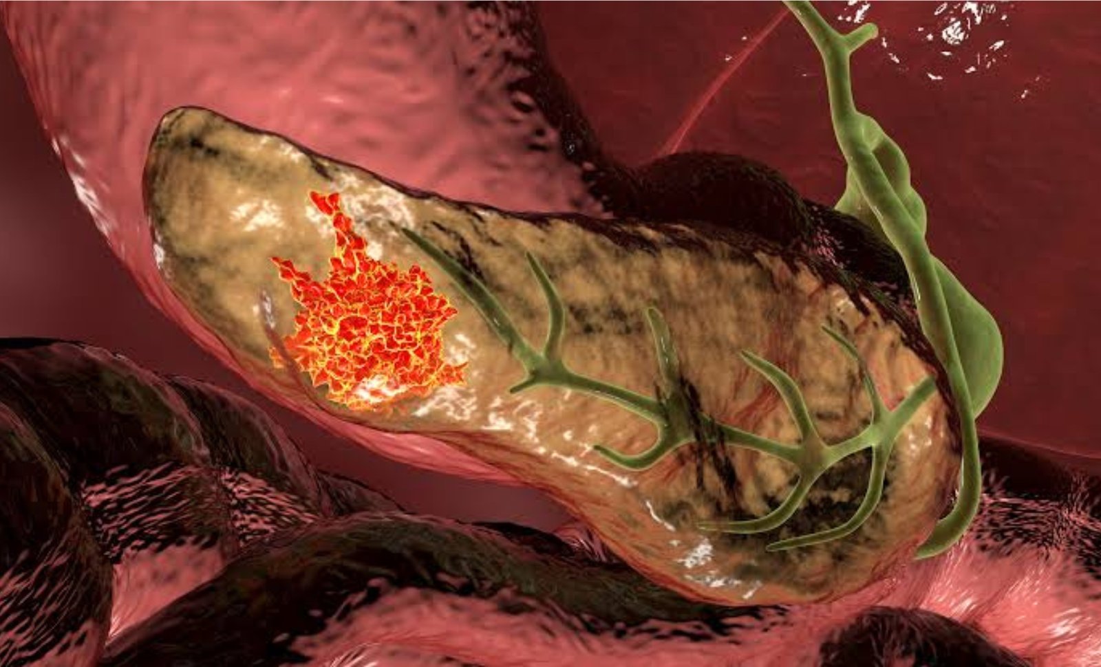
A CT scan can reveal a pancreas that looks perfectly normal, even when subtle changes associated with cancer are already developing. No visible tumor, no obvious warning signs, and nothing suspicious enough to trigger further investigation. Yet hidden within those same images may be clues to a disease that will not be diagnosed until months or years later.
An artificial intelligence system developed by researchers at Mayo Clinic and collaborating institutions has demonstrated an ability to detect those hidden signals. Called REDMOD, the technology identified imaging patterns associated with pancreatic cancer a median of 475 days before diagnosis, achieving nearly twice the sensitivity of experienced radiologists.
The findings, published in the medical journal Gut in April 2026 and highlighted in an October 26 report, suggest that existing medical scans may contain more information about developing pancreatic cancer than conventional visual interpretation can reveal.
The AI Found Warning Signs in Scans That Looked Normal
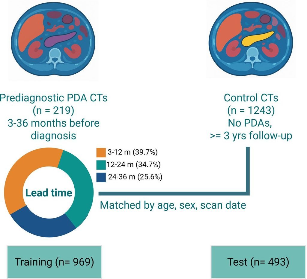
The investigation involved 1,462 abdominal CT scans, including 219 from patients who eventually developed pancreatic ductal adenocarcinoma, the most common form of pancreatic cancer.
At the time of their original examinations, those patients had no suspicious pancreatic masses or other obvious imaging abnormalities. Their scans had been taken between three months and three years before cancer was diagnosed.
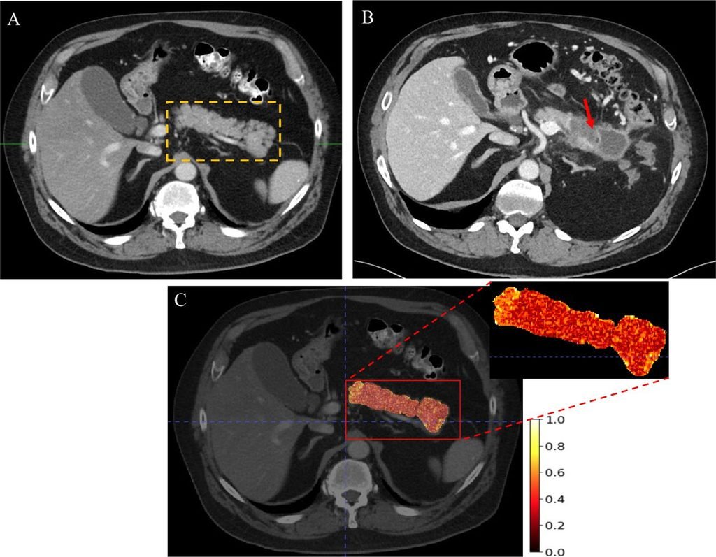
Another 1,243 scans came from people who remained free of pancreatic cancer during at least three years of follow-up.
After training REDMOD on 969 scans, the researchers evaluated its performance using 493 previously unseen images. Among 63 patients who later developed pancreatic cancer, the system correctly identified 46 cases, achieving a sensitivity of 73%.
Experienced radiologists evaluating the same scans achieved a combined sensitivity of approximately 38.9%.
But the most striking difference emerged in images taken furthest from diagnosis.
Its Advantage Was Greatest More Than Two Years Before Diagnosis
Pancreatic cancer is notoriously difficult to detect early. The pancreas lies deep within the abdomen, while developing tumors may produce few symptoms and little visible distortion of surrounding tissue.
REDMOD’s performance suggested that recognizable imaging patterns could emerge long before conventional abnormalities become apparent.
In scans taken three to twelve months before diagnosis, the AI identified 75% of future cancer cases, compared with 50% for radiologists. Between twelve and twenty-four months, the system maintained 75% sensitivity while radiologists reached approximately 41%.
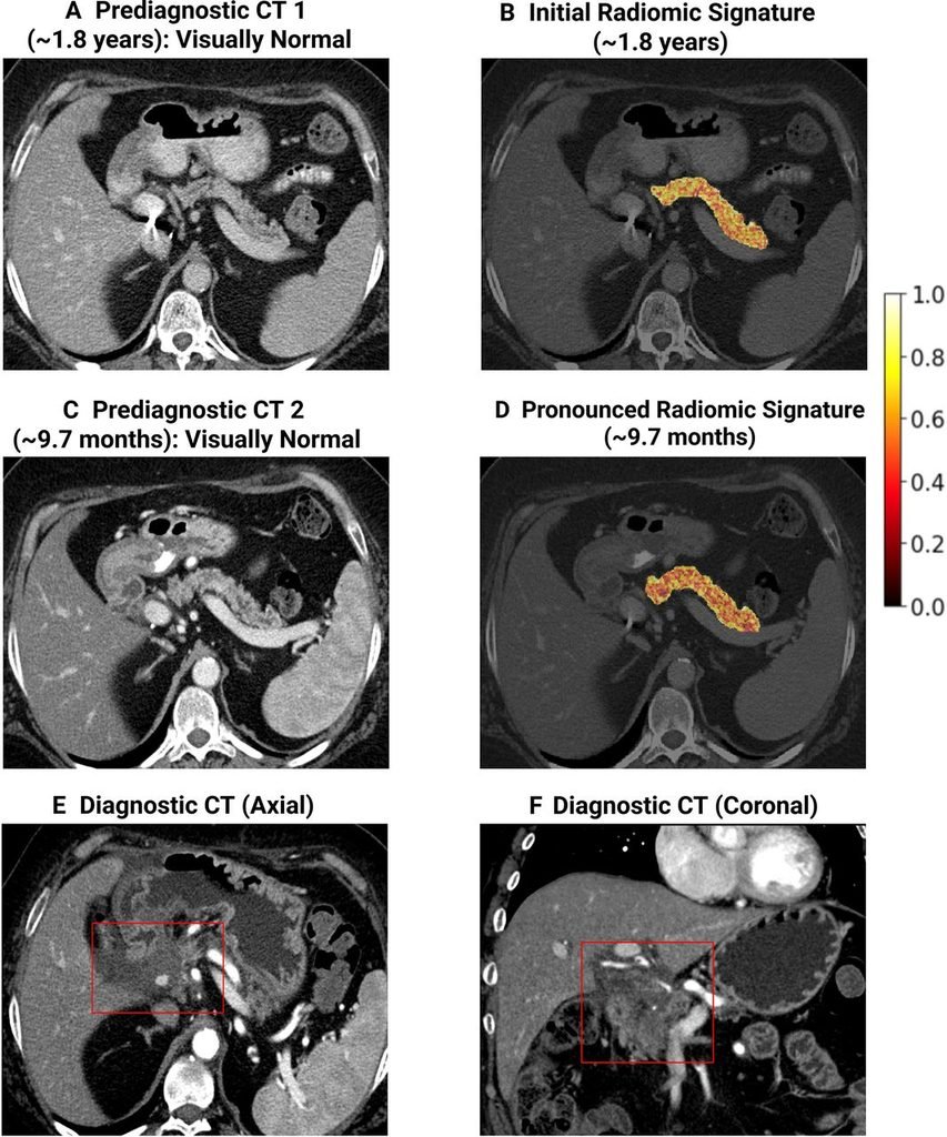
For scans obtained more than two years before diagnosis, REDMOD detected 68% of future cases, compared with just 23% for radiologists.
That represented almost three times the sensitivity at the earliest interval examined, although the relatively small subgroups limit the precision of those comparisons.
The system was not simply locating tumors that doctors had overlooked. It was measuring tissue characteristics that were difficult to distinguish through ordinary visual examination.
Forty Hidden Features Exposed Changes Invisible to Human Eyes
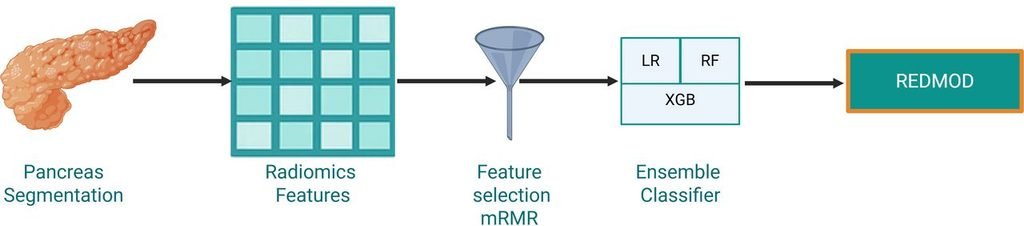
REDMOD uses a technique called radiomics, which transforms medical images into numerical measurements of tissue structure, intensity, and texture.
Instead of relying on an obvious mass, the system examines patterns distributed across pancreatic tissue. It automatically identifies the organ, extracts hundreds of imaging characteristics, and combines predictions from three machine-learning methods.
From an initial collection of 968 radiomic measurements, the researchers selected 40 features for the final model.
Remarkably, 36 came from images processed using wavelet filters, mathematical transformations that reveal subtle structural differences at multiple scales.
Those processed features substantially improved performance. A model using them achieved an area under the receiver operating characteristic curve, or AUC, of 0.82, compared with 0.74 for a model relying only on unfiltered features.
The original study published in Gut also documented how some suspicious imaging signatures persisted across multiple scans taken before diagnosis.
In one case, earlier images contained an abnormal radiomic signal in the region where a pancreatic tumor eventually became visible. The underlying changes had been detectable long before conventional imaging revealed a recognizable mass.
Earlier Detection Could Make a Critical Difference
Pancreatic cancer remains among the deadliest common malignancies, largely because it is frequently diagnosed after spreading beyond the pancreas.
The United States is expected to record approximately 67,530 new pancreatic cancer cases and 52,740 deaths in 2026.
According to National Cancer Institute survival statistics, five-year relative survival is approximately 43.6% when the disease remains localized. That figure drops to around 3.4% once cancer has spread to distant organs.
These differences make earlier detection a major priority.
A system capable of examining CT scans already obtained for other medical reasons could eventually help identify people who need closer investigation, particularly those with elevated pancreatic cancer risk.
However, detecting an early imaging signature is not the same as confirming cancer or demonstrating that earlier treatment will improve survival.
False Alarms Remain the Biggest Challenge
REDMOD’s higher sensitivity comes with a significant trade-off.
In its independent test set, the system correctly classified 349 of 430 cancer-free scans, producing a specificity of approximately 81.1%. The other 81 were incorrectly flagged as suspicious.
Applied to large populations, that false-positive rate could lead to substantial numbers of unnecessary tests and medical investigations.

The study also contained a much higher proportion of future cancer patients than routine screening would encounter, meaning its reported performance cannot be translated directly into general-population screening accuracy.
Further prospective testing is needed to determine whether doctors can use these alerts safely and whether identifying abnormalities earlier actually improves patient outcomes.
REDMOD is therefore a promising research tool, not an established pancreatic cancer screening test.
Still, its most remarkable finding lies in scans that initially appeared unremarkable: subtle pancreatic tissue patterns associated with future cancer were detectable more than two years before some patients received their diagnoses.

Farid Amber is a Canada-based writer and Editor-in-Chief who covers technology, space, and the environment. With a strong technical background, he brings a clear and practical perspective to stories about innovation, science, and the future of our planet.
His education includes a diploma in Applied Science, a master’s degree in Industrial Electrical Engineering, and a diploma in Electromechanical Automated Systems. This gives him a deep understanding of how modern technologies work, from complex engineering systems to the ideas driving new discoveries.
As Editor-in-Chief, Farid focuses on making complex topics easy to read, accurate, and meaningful. His goal is to help readers understand not only what is happening in the world of science and technology, but why it matters.
Leave a Reply