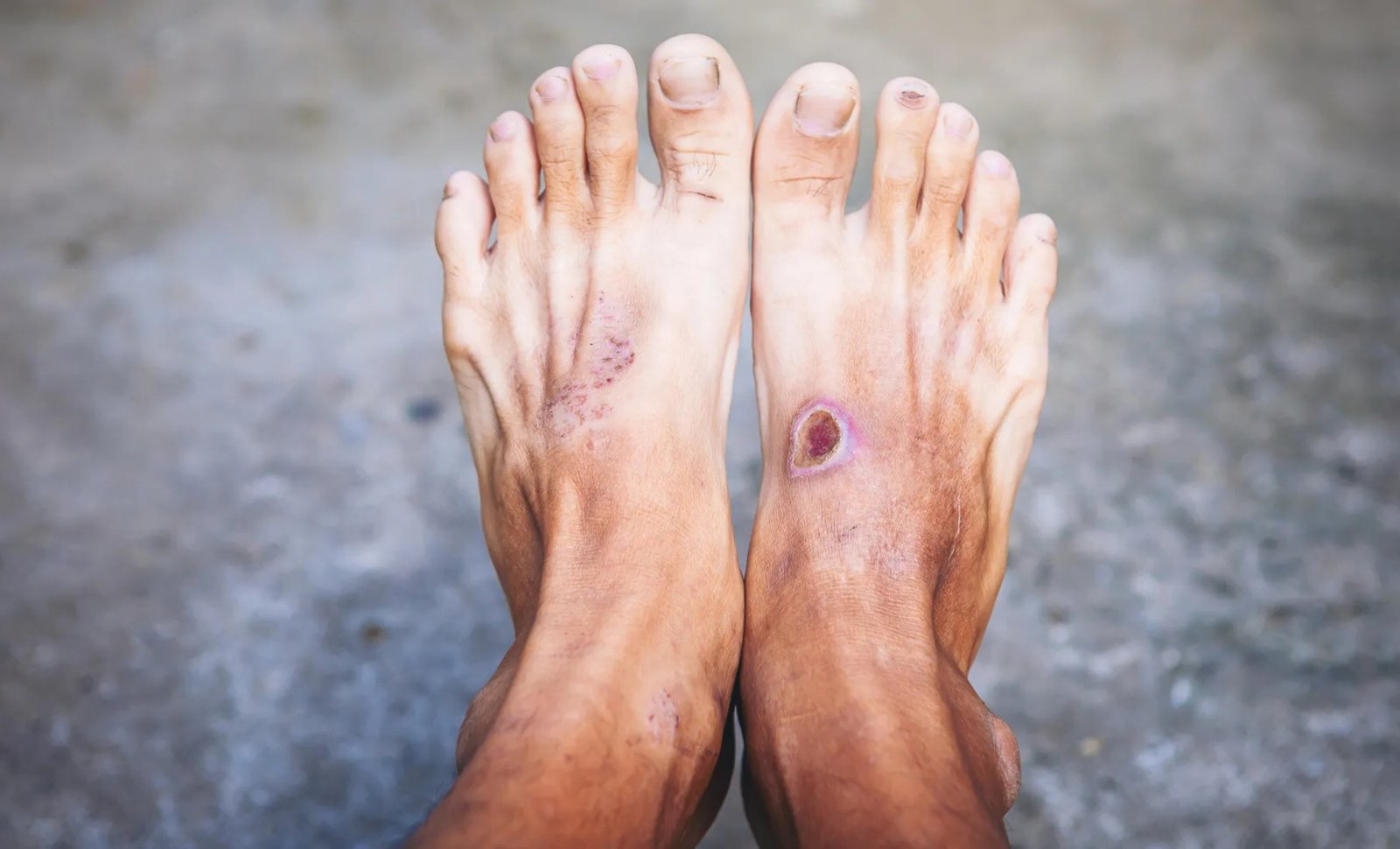
A small cut on the foot might heal within days for a healthy person. But for someone with diabetes, that same injury can develop into a wound that remains open for months. In severe cases, the damage can lead to infection, hospitalization or even amputation.
Doctors have long known that diabetes can interfere with the body’s ability to repair damaged tissue. Poor circulation, nerve damage and high blood sugar all contribute to the problem. Now, researchers in China are drawing attention to another possible culprit: immune cells that fail to switch from fighting inflammation to helping wounds heal.
In a review published in Frontiers of Medicine, scientists examined how different immune cells behave as diabetic wounds develop. Their analysis suggests that some cells remain unusually active long after their protective work should have ended, creating conditions that make tissue repair more difficult.
The research, led by Yi Ru and colleagues from Shanghai Skin Disease Hospital and other institutions in China, brings together earlier findings about eight groups of immune cells and how their behavior changes during wound healing.
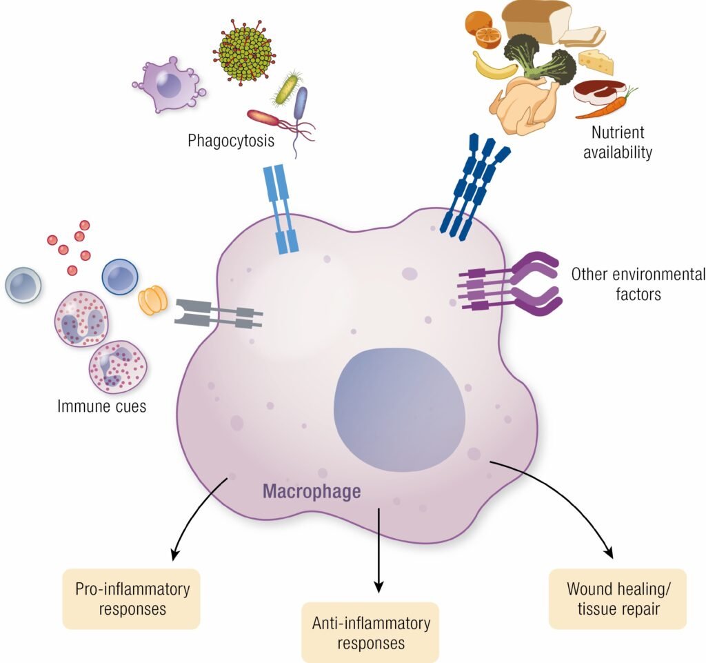
Among the most important are macrophages, immune cells responsible for clearing damaged tissue and helping coordinate the body’s response to injury.
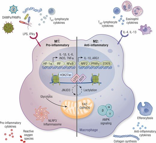
When a wound first appears, macrophages often take on an inflammatory role, helping defend the injured area against microorganisms. As healing progresses, their functions normally shift toward supporting tissue repair, allowing new skin and blood vessels to form.
Scientists have traditionally described these two broad patterns as M1 and M2 macrophage states. The first is associated with inflammation, while the second is more closely linked to repair.
In people with diabetes, however, macrophages can remain in inflammatory states for too long. Instead of helping the wound advance toward recovery, they continue releasing signals that can contribute to tissue damage.
The researchers also caution that the M1/M2 distinction is an oversimplification. Macrophages can adopt a wider range of behaviors, making it difficult to divide them neatly into cells that damage tissue and cells that repair it.
And macrophages are not the only immune cells involved.
Neutrophils, which are among the first cells to reach an injury, can also contribute to chronic inflammation. They normally help defend wounds by attacking microorganisms and releasing web-like structures known as neutrophil extracellular traps, or NETs.
But these defenses can become poorly regulated in diabetic wounds. Excessive NET formation, together with the release of damaging enzymes and reactive oxygen species, can injure surrounding tissue and prolong inflammation.
Another problem may arise when the immune system attempts to remove dead cells.
During normal healing, damaged cells are cleared away through a process called efferocytosis. Certain immune cells, including dendritic cells, help carry out this cleanup. When it works properly, the removal of dead cells helps inflammation subside.
Evidence reviewed by the researchers suggests that dendritic cells may become less effective at this task in diabetic wounds. As dying cells accumulate, they can contribute to continued inflammatory signaling.
The researchers identified a transporter called SLC7A11 as one molecular factor involved in this process. Reduced activity of the transporter under diabetic conditions may impair dendritic cell function and interfere with the normal resolution of inflammation.
Other immune cells appear to suffer disruptions of their own. Regulatory T cells, which help prevent excessive immune responses, can become less numerous or less effective in diabetic wounds. Specialized T cells in the skin may also produce fewer of the growth signals needed to regenerate damaged tissue.
Some of these cells normally help stimulate keratinocytes, which make up much of the skin’s outer layer. Without sufficient support, the process of covering a wound with new skin can slow down.
The review also examines B cells and natural killer cells, two immune populations whose roles in diabetic wound healing remain less thoroughly understood. Emerging research suggests that B cells may help influence macrophage behavior, while natural killer cells can affect inflammatory signaling and blood vessel formation.
Taken together, the findings suggest that chronic diabetic wounds involve more than a single malfunctioning immune cell. Several parts of the immune system may fail to coordinate their activities as an injury moves from inflammation toward repair.
That possibility has prompted scientists to investigate treatments designed to change how immune cells behave inside wounds.
Some experimental approaches seek to encourage macrophages to adopt more repair-associated functions. Others use biological drugs to interrupt inflammatory signals that remain active for too long.
The researchers also highlight mesenchymal stem cells and tiny particles released by these cells, known as extracellular vesicles. Both have attracted interest for their potential to influence inflammation and tissue regeneration.
Specialized wound dressings could eventually offer another way to deliver these treatments. Rather than exposing the entire body to an immune-modifying substance, advanced biomaterials may allow therapeutic agents to be released directly into injured tissue.
Several other approaches are under investigation, including compounds that regulate oxidative stress and treatments intended to stimulate particular immune cell populations.
Still, altering immune activity is not necessarily as simple as reducing inflammation. The body’s initial inflammatory response is essential for fighting infection and clearing damaged tissue. Suppressing that response at the wrong time could interfere with healing rather than improve it.
The researchers therefore emphasize the importance of timing.
Wounds move through overlapping stages of inflammation, tissue growth and remodeling. Different immune cells become more or less active throughout those stages, meaning a treatment that helps during one phase might be less useful during another.
Future strategies could involve examining the immune cells present in an individual wound before choosing a treatment. Such an approach might help clinicians identify whether a wound needs stronger control of inflammation or additional support for tissue regeneration.
For now, the research offers a framework for future treatment development rather than a new cure. The paper is a review of existing scientific evidence, not a clinical trial demonstrating that a particular immune-targeting therapy can reliably heal diabetic ulcers.
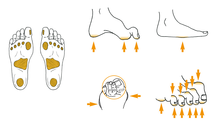
Established wound care remains essential. The International Working Group on the Diabetic Foot’s practical guidelines recommend reducing pressure on affected areas, assessing blood supply and addressing infection when necessary. For certain ulcers on the soles of the feet, the group’s offloading recommendations identify specialized devices that keep pressure off the wound as a first-choice treatment.
The scientists’ longer-term goal is to understand how these established treatments might be complemented by therapies that address the immune dysfunction occurring beneath the surface.
One particularly important finding is that immune cells do not simply become helpful or harmful in isolation. Their functions change as a wound progresses, and the signals they exchange can determine whether inflammation continues or tissue repair moves forward.

Ghiles Houari is a writer based in Algeria with a technical background in applied science and industrial electronics. He is naturally curious about how things work, from everyday technology to the systems and ideas shaping the modern world.
With his hands-on technical training, Ghiles brings a practical and thoughtful perspective to his writing. He focuses on making science, technology, electronics, and innovation clear, interesting, and easy to understand.
For him, good writing is not just about explaining facts — it is about helping readers see why they matter.
Leave a Reply